USG (Ultrasound)-guided biopsy
An ultrasound-guided biopsy is a minimally invasive diagnostic procedure that uses real-time ultrasound imaging to guide the extraction of tissue samples from suspicious or abnormal areas within the body. This technique is widely used to evaluate lumps, masses, or lesions detected during physical exams or imaging studies. It is particularly effective for superficial or soft tissue structures, such as the breast, thyroid, liver, lymph nodes, or muscles.
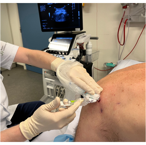
Procedure:
The patient is positioned appropriately, and a local anesthetic is applied to the biopsy site to minimize discomfort.
A handheld ultrasound probe is used to generate real-time images of the target area, allowing the physician to visualize the abnormality and surrounding structures.
Using the ultrasound images as a guide, a fine needle or core biopsy needle is inserted into the target area to collect a tissue sample.
The sample is sent to a laboratory for histopathological analysis to determine the nature of the abnormality (e.g., cancerous, benign, or infectious).
Advantages:
Real-time imaging: Provides continuous visualization, ensuring precise needle placement.
Non-ionizing radiation: Unlike CT or X-rays, ultrasound uses sound waves, making it safer for repeated use.
Minimally invasive: Reduces the risk of complications compared to surgical biopsies.
Cost-effective: Generally less expensive than other imaging-guided biopsy techniques.
Common Uses:
Diagnosing breast lumps or abnormalities.
Evaluating thyroid nodules.
Assessing liver lesions or tumors.
Investigating suspicious lymph nodes or soft tissue masses.
Risks:
Mild pain, bruising, or bleeding at the biopsy site.
Rare complications, such as infection or damage to nearby structures.
Ultrasound-guided biopsies are a safe, accurate, and widely used diagnostic tool, providing critical information for diagnosing and managing various medical conditions.