Best Diagnostic Centre in Chennai
Providing Quality and affordable service since 1987
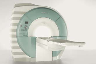
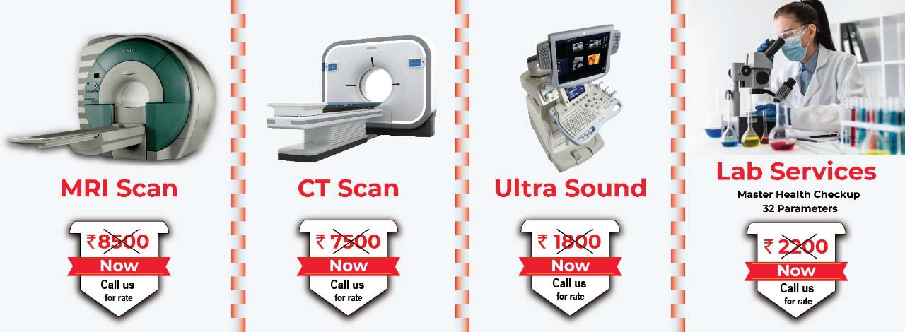
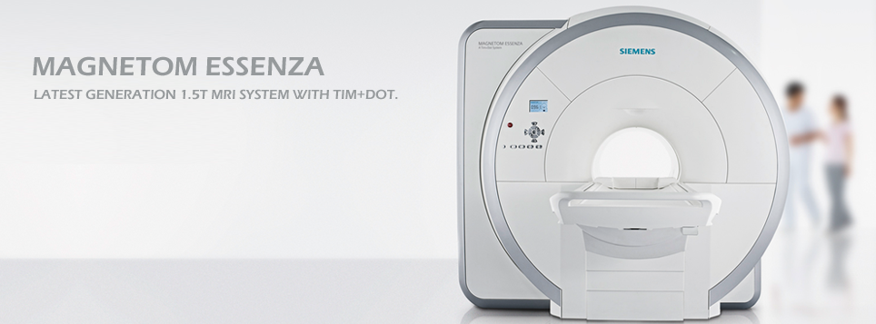
Magnetic resonance imaging (MRI) is a test that uses a magnetic field and pulses of radio wave energy to make pictures of organs and structures.MRI Scan in Chennai
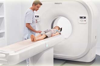
Computed Tomography (CT) can be used to help diagnose, and plan the treatment, for a range of medical conditions. CT scan in Chennai
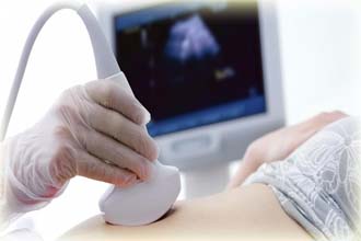
Ultrasound scanning or sonography, involves exposing part of the body to high-frequency sound waves to produce pictures of the inside of the body. Ultrasound Scan in chennai
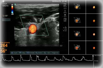
Color Doppler ultrasound is a medical imaging technique which is used to provide visualization of the bloodflow can clearly see what is happening inside the body.
The electroencephalogram (EEG) is a measure of brain waves. It is a readily available test that provides evidence of how the brain functions over time.
Digital X-Ray Mammogram and High definition sono mammogram with excellent image Quality for accurate diagnosis. With excellent image quality for accurate diagnosis. X Ray Mammogram in Chennai
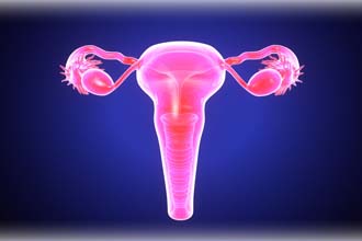
HSG is an important test of female fertility potential. The HSG test is a radiology procedure usually done in the radiology department of a hospital or outpatient radiology facility. HSG Scan in Chennai
X-ray picture is really a picture of the shadows cast by the denser materials (like bones) in your body
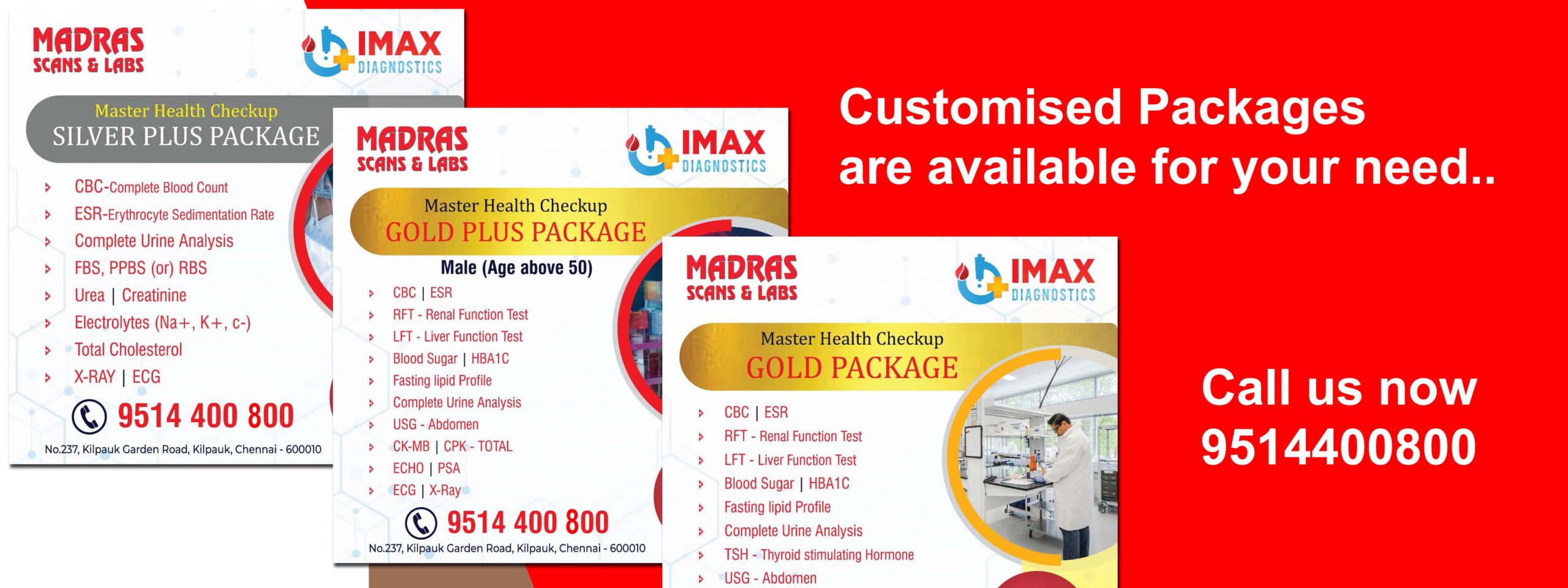
Lab tests are critical tools in helping healthcare providers to understand what is happening in your body. The test results can help confirm the cause of health concerns or rule out any potential concerns.
Importance of Diagnostic Services
Diagnostic services play a crucial role in the healthcare industry by helping to identify, diagnose, and monitor various medical conditions. With advancements in medical technology and increasing awareness about healthcare, the demand for diagnostic services has been growing rapidly. Here are some of the best diagnostic services available today:
Radiology and Imaging Services – Radiology and Imaging services are used to diagnose and monitor a variety of medical conditions using various imaging techniques such as X-rays, CT scans, MRI scans, ultrasound. Some of the best radiology and imaging services providers include Madras Scans & Labs and Radiology Partners.
Clinical Laboratory Services – Clinical laboratory services include a wide range of tests and procedures that are used to diagnose and monitor various medical conditions. These tests may include blood tests, urine tests, genetic tests, and other specialized tests. Some of the best radiology and imaging services providers include Madras Scans & Labs and Laboratory Partners.
Pathology Services – Pathology services are used to diagnose and monitor various medical conditions by analyzing tissue samples and other specimens. These services are particularly useful in the diagnosis of cancer and other diseases. Some of the best radiology and imaging services providers include Madras Scans & Labs and Pathology Service Partners.
Cardiology Services – Cardiology services are used to diagnose and treat heart-related conditions such as heart disease, heart attacks, and arrhythmias. These services may include electrocardiograms (ECGs), stress tests, echocardiograms, and other specialized tests. Some of the best cardiology services providers include Madras Scans & Labs and Partners.
Neurology Services – Neurology services are used to diagnose and treat conditions that affect the nervous system, such as epilepsy, multiple sclerosis, and Parkinson’s disease. These services may include electroencephalograms (EEGs), nerve conduction studies, and other specialized tests. Some of the best neurology services providers include Madras Scans & Labs and Partners.
Overall, the best diagnostic services providers are those that offer accurate and timely results, use state-of-the-art technology, and have experienced and qualified staff. We at Madras Scans take care of everything to feel comfort. It’s essential to choose a provider that has a good reputation in the healthcare industry and offers a wide range of services to meet your healthcare needs.
For a Best Diagnostic service in Chennai, consider Madras Scans & Labs as your go-to destination. It is the most trusted center with its exceptional and secure diagnostic services at an affordable pricing system.
Give us a quick call at 9514400800 or visit us at https://www.madrasscan.in to know more about us and our attractive price package.
Call us for an appointment
(+91) 9514400 800
Feel free to contact us.
admin@madrasscan.in
Reach to us via our location
View Map

