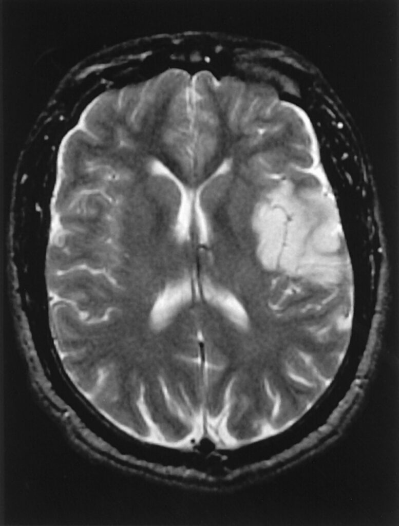
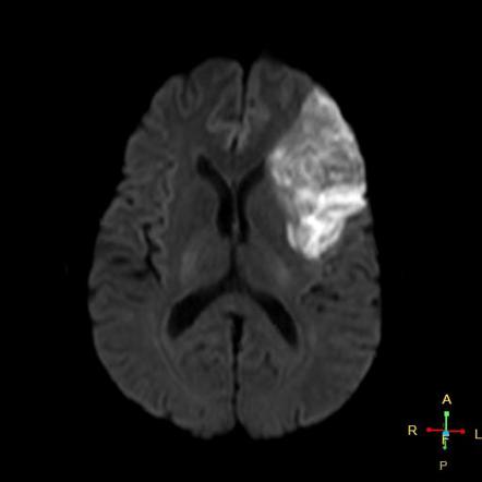
MRI (Magnetic Resonance Imaging) is a commonly used diagnostic tool for brain strokes. MRI is a non-invasive imaging technique that uses a strong magnetic field and radio waves to create detailed images of the brain. It can detect the presence, location, and extent of brain damage caused by a stroke.
When a stroke occurs, brain cells begin to die due to a lack of oxygen and nutrients. An MRI can detect this damage by producing images that show the affected areas of the brain. The MRI can also show the type of stroke, whether it is caused by a blood clot or bleeding in the brain.
An MRI scan can be performed with or without contrast. Contrast is a special dye that is injected into the body before the MRI to make certain tissues or blood vessels more visible. Contrast can help to identify the location and severity of the stroke.
In addition to detecting the presence of a stroke, an MRI can also help doctors assess the extent of brain damage and determine the appropriate treatment. MRI scans are often used to monitor the progress of stroke patients and to assess the effectiveness of treatments over time.
Overall, MRI scans are a valuable tool in the diagnosis and management of stroke. They provide detailed information about the location and severity of brain damage, which can help doctors make more informed treatment decisions and improve outcomes for stroke patients.