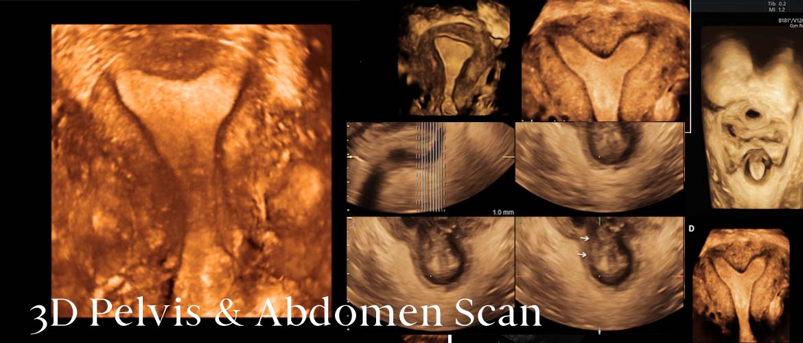
3D Pelvis & Abdomen Scan in Chennai
Special Features:
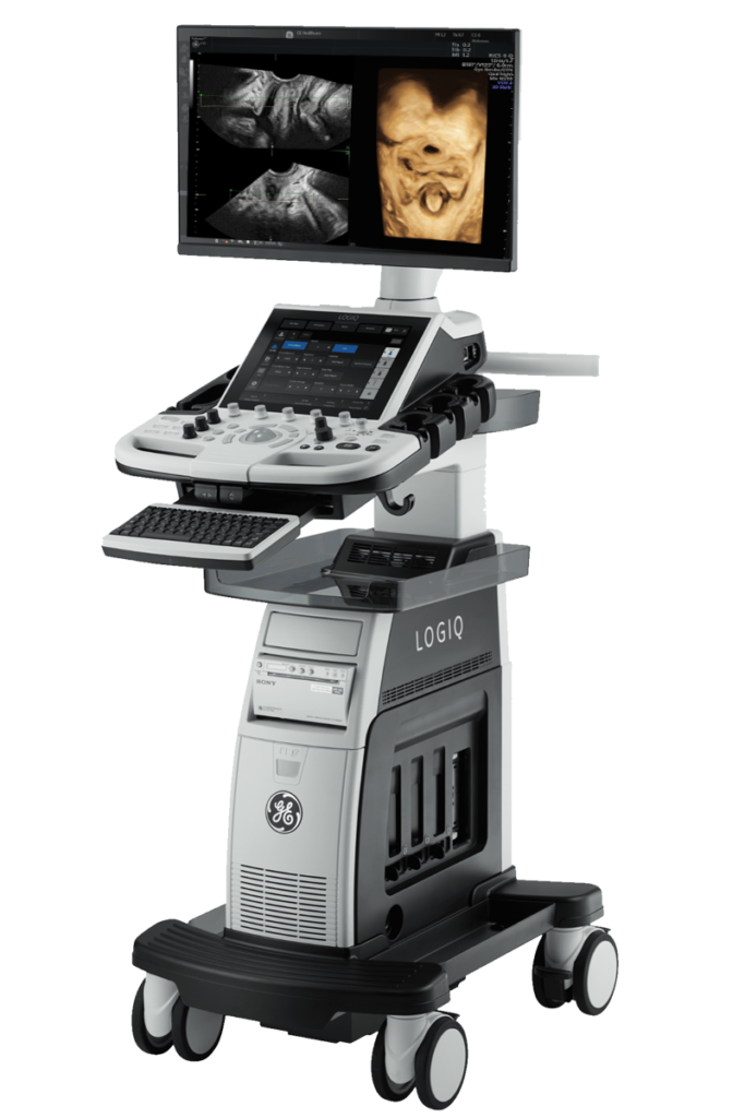
SCAN DONE BY LATEST GE LOGIC P10 USG MACHINE
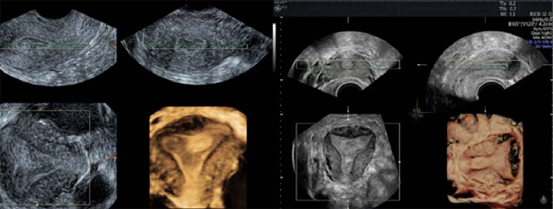
3 DIMENSIONAL VIEW OF PELVIS
SCAN DONE ON ALL DAYS
EXCELLENT IMAGING, ACCURATE RESULTS
QUICK REPORTING
Advantages of 3D Imaging over Routine Ultrasound:
Accuracy of the 3D images is 85-95% according to Journal of Ultrasound Study
Helps in Better Diagnosis
For Better Treatment Modalities by the Gynecologist
To assess Ovarian volume & Determine the Ovarian Flow
Better assessment of position and nature of uterine fibroids
In Ascertaining Thickness of Endometrial Cavity to rule out Hyperplasia
To provide better opportunities in Diagnosis and Treatment of Mullerian Duct Anomalies
Towards Better Solutions in IVF/Infertility
In assessing the Internal Characteristics of Ovarian Cysts
In co-ordinating with the Transvaginal Ultrasound in Follicular Counts
3D Abdomen ultrasound Scan in Chennai
A 3D abdomen ultrasound scan is a medical imaging technique that uses sound waves to create three-dimensional images of the abdominal organs. This technology is an advancement over traditional 2D ultrasound, providing additional information and benefits in various medical scenarios.
Some of the advantages of 3D abdomen ultrasound scans:
Improved Visualization: 3D ultrasound provides a more detailed and realistic representation of the abdominal organs compared to 2D imaging. This can be especially useful for visualizing complex structures and abnormalities.
Diagnostic Accuracy: The enhanced visualization can lead to increased diagnostic accuracy, helping healthcare professionals identify and assess various conditions affecting the abdomen, such as tumors, cysts, and other abnormalities.
Fetal Imaging: In obstetrics, 3D ultrasound is commonly used for fetal imaging. It allows for detailed visualization of the developing fetus, providing valuable information about fetal anatomy and development. Parents may also benefit from the enhanced imaging quality for bonding and emotional reasons.
Guidance for Procedures: 3D ultrasound can assist in guiding medical procedures such as biopsies or drainage of fluid collections. The real-time imaging helps healthcare providers navigate through the targeted area more precisely.
Surgical Planning: For certain abdominal surgeries, preoperative 3D ultrasound images can aid surgeons in planning procedures by providing a comprehensive view of the affected organs and surrounding structures.
Monitoring and Follow-up: 3D ultrasound can be used to monitor changes in abdominal organs over time, making it valuable for follow-up examinations and tracking the progression or regression of certain conditions.
Reduced Operator Dependency: 3D ultrasound technology may reduce the dependency on the operator’s skill in obtaining quality images compared to traditional 2D ultrasound. This can lead to more consistent and reproducible results.