Low Cost CT Brain Scan and its uses
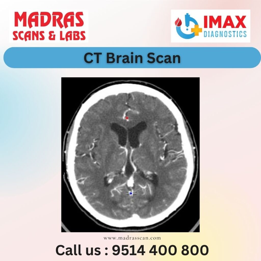
A CT (computed tomography) brain scan, also known as a CT head scan, is a medical imaging technique that uses X-rays and computer processing to create detailed cross-sectional images of the brain. CT scans are valuable diagnostic tools for assessing various conditions affecting the brain, including injuries, tumors, infections, bleeding, and other abnormalities. Here are some common uses and procedures associated with CT brain scans:
Uses:
Traumatic Brain Injury (TBI): CT scans are frequently used to evaluate head injuries and assess the extent of damage caused by trauma.
Stroke: CT scans can help identify areas of the brain affected by stroke, whether due to ischemia (lack of blood flow) or hemorrhage (bleeding).
Tumors: CT scans are useful in detecting and characterizing brain tumors, providing information about their size, location, and characteristics.
Infections: Brain infections, such as abscesses or encephalitis, can be visualized through CT scans.
Vascular Conditions: CT angiography (CTA) may be performed to assess blood vessels in the brain, identifying conditions like aneurysms or arteriovenous malformations.
Hydrocephalus: CT scans can help diagnose conditions where there is an abnormal accumulation of cerebrospinal fluid in the brain.
Degenerative Disorders: Some degenerative disorders, like Alzheimer’s disease, may show characteristic changes on CT scans, although other imaging modalities like MRI are often preferred for such conditions.
Procedure:
Patient Preparation: Before the procedure, the patient may be asked to remove any metal objects and may need to fast for a few hours, especially if contrast material will be used.
Contrast Material (Optional): In some cases, a contrast material (iodine-based dye) may be injected into a vein to enhance the visibility of certain structures or abnormalities.
Positioning: The patient is positioned on a movable table, which is then moved into the CT scanner.
Scanning: The CT scanner rotates around the patient, capturing X-ray images from various angles. The X-ray detector and the computer process the data to create detailed cross-sectional images (slices) of the brain.
Monitoring: The patient is monitored during the procedure, and the technologist may communicate with them through an intercom system.
Post-Processing: After the scan is complete, the images are reviewed by a radiologist to make a diagnosis.
CT brain scans are relatively quick and provide detailed images that can assist healthcare professionals in diagnosing and planning treatment for various neurological conditions. However, it’s important to note that ionizing radiation is involved in CT scans, and considerations about its use and potential risks are taken into account, especially in certain patient populations.
For a doing painless CT-Brain Scan in Chennai, consider Madras Scans & Labs as your go-to destination. It is the most trusted Scan center in Chennai with its exceptional and secure diagnostic services at an affordable pricing system.
Give us a quick call at 9514400800 or visit us at https://www.madrasscan.in to know more about other scans and our attractive price package.