Parkinson’s disease (PD) is a neurodegenerative disorder that primarily affects movement. MRI is a non-invasive imaging technique that can detect changes in brain structure and function in people with PD.
Early MRI findings in PD include:
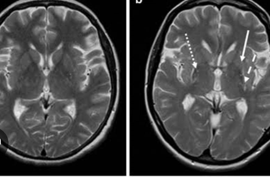
Decreased volume of the substantia nigra: This is a region in the brain that is responsible for producing dopamine, a neurotransmitter that is important for movement. A decrease in the volume of this region is a hallmark feature of PD and can be detected using MRI.
Increased iron accumulation in the substantia nigra: In PD, there is an abnormal accumulation of iron in the substantia nigra. This can also be detected using MRI.
Changes in the basal ganglia: The basal ganglia is a group of nuclei in the brain that are involved in controlling movement. In PD, there are changes in the basal ganglia that can be detected using MRI.
Thinning of the cerebral cortex: The cerebral cortex is the outer layer of the brain that is responsible for higher brain functions such as thinking and perception. In PD, there may be a thinning of the cerebral cortex, which can be detected using MRI.
It is important to note that these early MRI findings may not always be specific to PD and can also be seen in other neurodegenerative disorders. A diagnosis of PD is typically based on a combination of clinical symptoms and imaging findings.
For a painless MRI Scan Chennai, consider Madras Scans & Labs as your go-to destination. It is the most trusted MRI scan center in Chennai with its exceptional and secure diagnostic services at an affordable pricing system.
Give us a quick call at 9514400800 or visit us at https://www.madrasscan.in to know more about MRI Scan and our attractive price package.